
Pancreas, bile duct & gut wall
Endoscopic Ultrasound (EUS) in Delhi
Ultrasound from inside the gut, with biopsy and drainage in the same procedure
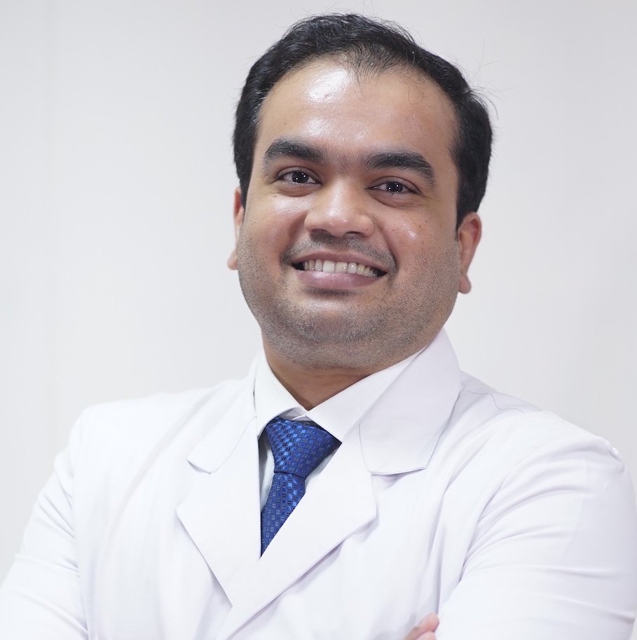
Dr Surakshith T K has performed more than 5,000 EUS procedures in Delhi, including EUS-guided biopsy and therapeutic EUS. EUS is used when CT and MRI cannot answer the question — a small pancreatic lesion, a duct stone that scans have missed, or a lump in the wall of the stomach.
What is endoscopic ultrasound (EUS)?
EUS combines an endoscope with an ultrasound probe at its tip, so the pancreas, bile duct, and the individual layers of the gut wall can be imaged from a few millimetres away rather than through the abdominal wall. That proximity gives detail CT and MRI cannot match for small lesions. The same procedure can take a needle biopsy (FNA or FNB) or place a stent to drain a collection, so diagnosis and treatment often happen together.
Indications
What EUS is used for
EUS answers questions about small structures and about depth — both of which decide treatment.
EUS underpins much of advanced endoscopy in Delhi: it tells you the depth before an ESD, the layer of origin before an EFTR, and the cause of obstruction before an ERCP.
The procedure
How EUS is performed
- Preparation — fasting before the procedure and a review of blood-thinning medication
- Sedation — deep sedation, occasionally general anaesthesia for therapeutic cases
- Imaging — the echoendoscope is passed to the stomach or duodenum and the area is scanned systematically
- Sampling — if a lesion needs tissue, a fine needle is passed through the scope under real-time ultrasound
- Therapy if planned — drainage or stent placement in the same sitting where that was the intention
- Afterwards — most diagnostic patients go home the same day once the sedation has worn off
What EUS can and cannot settle
A needle biopsy sometimes comes back inconclusive. That is a known limitation, not an error, and it occasionally means repeating the sampling.
EUS is operator-dependent to a degree that imaging is not. The value of the report depends on who performed the study and how systematically the area was scanned.
EUS complements CT and MRI rather than replacing them. The scans map the territory; EUS answers the specific question about a small structure.
Related
Related procedures
Been advised an EUS, or a biopsy of a pancreatic lesion?
Send your CT, MRI or MRCP and blood reports. EUS is often the step that turns a suspicious finding on a scan into a definite diagnosis, and it can sometimes be combined with treatment in the same sitting.
- +91 96861 37977
- Kalka Gastro Liver & Obesity Clinic10/22 Nehru Enclave East, Kalkaji, New Delhi 110019
- Mon–Sat, 5:00 pm – 8:30 pmProcedures at Fortis Escorts (Okhla) & Holy Family (NFC)
FAQ
Frequently asked questions
Is EUS better than a CT or MRI scan?
They answer different questions. CT and MRI show the whole abdomen and are the right first tests. EUS images small structures — the pancreas, the bile duct, the layers of the gut wall — from a few millimetres away, so it detects and characterises lesions that scans cannot resolve, and it can take a biopsy at the same time. It is used to answer a specific question, not as a general screening test.
Is EUS painful?
No. EUS is performed under sedation and patients are asleep for it. Most people have nothing more than a sore throat and some bloating afterwards, and diagnostic patients usually go home the same day.
Is an EUS-guided biopsy safe?
Yes. The needle passes only through the wall of the stomach or duodenum and travels a very short distance under continuous ultrasound vision, with Doppler used to avoid blood vessels. For pancreatic lesions it is the preferred route for obtaining tissue.
- MBBS · MD (Medicine) · DNB (Gastroenterology & Hepatology), Sir Ganga Ram Hospital
- Senior Consultant Gastroenterologist, Hepatologist & Therapeutic Endoscopist
- 250+ third-space procedures · 200+ POEM · 10,000+ ERCP · 5,000+ EUS
- Fortis Escorts Hospital, Okhla · Holy Family Hospital, New Delhi · Kalka Gastro Liver & Obesity Clinic, Kalkaji
- Delhi Medical Council reg. 74686
This page is for general information and does not replace an individual consultation. Whether a procedure is suitable depends on your own reports and examination. Last reviewed September 2026.